Treatment

What Is Macular Degeneration?

Macular Degeneration can lead to severe vision loss and occurs in the central part of the retina, the macular. It is most common in those over 60 however in some rare cases can occur at a younger age. It often occurs in both eyes however the progression in each eye can differ. The degeneration occurs in the retina of the eye so the peripheral vision is not effected therefore will not lead to complete blindness.

Symptoms:

- Text appears blurry making it difficult to read

- Difficulty to recognise faces

- Colours do not appear as vibrant

Forms of Macular Degeneration:

Dry: The dry form of macular degeneration is when there are yellow deposits called drusen in the macular. As these grow and increase this is what can lead to the dimming of vision. This is often most apparent when reading. Blind spots in the center of the persons vision can also occur as well as the loss of central vision in severe cases.

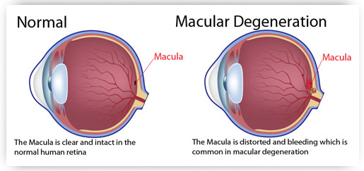
Wet: The wet form of macular degeneration is when there is a growth of abnormal blood vessels underneath the macular. The vessel begin to leak blood and fluid into the retina which is what causes the distortion of vision. Side effects include straight lines appearing wavy and blind spots which give a loss of vision. Eventually scarring can happen which will lead to permanent vision loss.

Dry macular degeneration is the most common out of the two forms, however dry macular degeneration can lead to the wet form. Research has found only 10% will develop the wet form and these make up for the majority of those who will suffer from serious vision loss.

Macular degeneration can change over time so it is key that those who suffer from either form monitor their eyesight and have regular eye examinations. 

3D OCT Screening Diagnoses

What is OCT?

OCT – Ocular Coherence Tomography – is an advanced eye scan for people of all ages. Similar to an ultrasound, OCT uses light rather than sound waves to illustrate the different layers that make up the back of the eye. The OCT machine captures both a fundus photograph and a cross-sectional scan of the back of the eye at the same time.

It is a new, completely painless and highly advanced screening system that checks for potentially serious conditions such as glaucoma, diabetes, age-related macular degeneration, vitreous detachments and more.

Your optometrist will take both a digital photograph and a three dimensional cross sectional scan of the back of your eye in one sitting. The scan is non-invasive, painless, simple and quick.

Step 1 – Book an appointment with your optometrist.

Step 2 – The optometrist will scan your eyes using the 3D OCT camera.

Step 3 – The high resolution 3D images are examined by the optometrist using specialist built-in analysis tools.

Step 4 – The results are presented to you.

Step 5 – Any future scans can be compared with previous ones for comparative diagnosis.

What can the scan check for?

1. Age Related Macular Degeneration

OCT can identify this condition and it's type (both wet and dry) and also monitor its progress, for example if you are undergoing treatment for sure a condition.

2. Diabetes

In the UK, more than two million people have been identified as having diabetes. OCT examination enables early detection, which greatly improves the success rate of treatment.

3. Glaucome

Around two in every 100 people over the age of 40 are affected by some form of glaucoma. The danger with chronic glaucoma is that there is no pain and your eyesight will seem to be unchanged, but your vision is being damaged. An OCT examination will confirm if you are at risk or what stage of glaucoma you may have.

4. Macular Holes

A small hole In the macular – the part of the retina which is responsible for our sharp, detailed, central vision. There are many causes of macular holes, one is caused by vitreous detachment, when the vitreous pulls away from the back of the eye and sometimes it does not 'let go' and eventually tears the retina, leaving a hole. Extreme exposure to sunlight can also be another cause.

5. Vitreous Detachments

As people get older the vitreous jelly that takes up the space in our eyeball can change. It can become less firm and move away from the back of the eye towards the centre, in some cases parts do not detach cause 'pulling' of the retinal surface. The danger of this detachment is that there is no pain and your eyesight may remain the same but the back of the eye could be damaged.

What does OCT cost?

There may be an additional charge of the OCT scan, but the benefits are obvious. You can enjoy the peace of mind knowing your eyes are in great condition.